8 Feb, 2022
Ultrasound Scans: The Complete Guide
Ultrasound Scans: The Complete Guide
If you've never had an ultrasound scan before, you might be wondering how they work, and what you can expect from your procedure. You can rest assured that ultrasound scans are safe and widely used. This definitive guide to ultrasounds will answer any questions you might have before your scan, including how ultrasound scans work, what they can show, how to prepare for an ultrasound, and how to book a private ultrasound scan.
What is an ultrasound scan?
An ultrasound scan is a medical imaging technique, which enables external and internal scanning of organs, soft tissues, the musculoskeletal system (including muscles, tendons, ligaments and joints) and the vascular (circulatory) system.
Ultrasound scans provide pictures (sonograms) of the inside of your body, allowing diagnosis of certain medical conditions (sonography), an understanding of how your body is working, feedback on the progress of some treatments, and to view a developing foetus during pregnancy. Ultrasounds can also capture real-time images to show how body parts are working in motion.
Ultrasound scans were first used in clinical practice in 1956, and are one of the most popular medical imaging techniques available today.
How do ultrasound scans work?
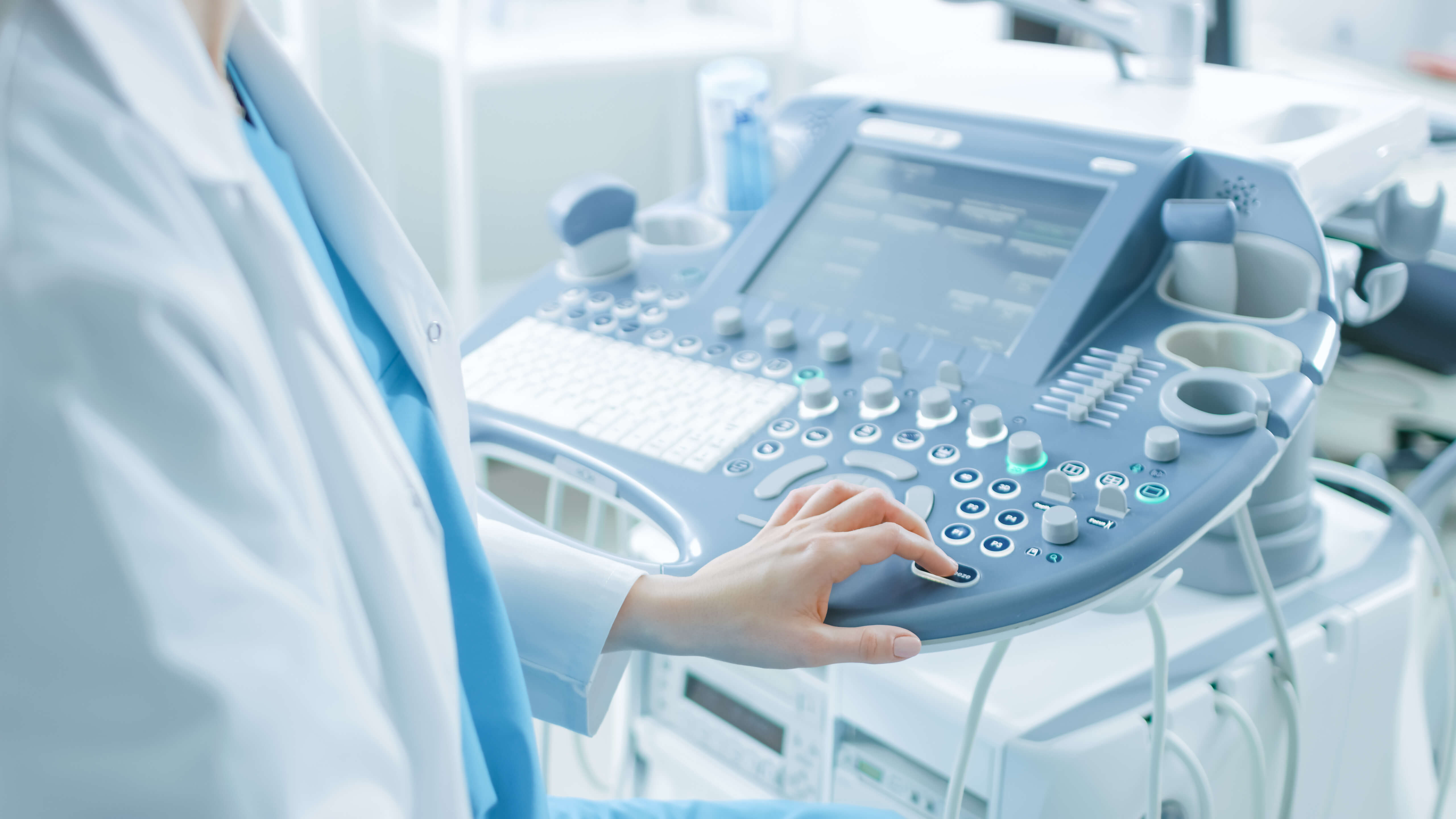
Ultrasound imaging is also known as sonography because it uses high-frequency sound waves to capture images inside the body in real-time.
An ultrasound probe is called a transducer, and there are different types and sizes that can be used either on the skin (externally) or within the body (internally).
The probe emits the ultrasound waves, which are reflected by soft tissues, fluid or bone within the body. The transducer then records the speed and timing of these echoes to generate real-time images, which can be viewed on a monitor during the scan.
💡 Humans can generally hear sounds up to 20KHz pitch, with dogs and cats able to hear up to 40KHz. Most ultrasound scans use much higher frequencies in the megahertz (MHz) range, meaning they are so high-pitched they cannot be heard by humans.
Ultrasounds are not always suited to organs that contain air, such as the stomach, lungs and bowels, because the ultrasound waves do not bounce off the structures very well. This means the echoes are harder to pick up and clear images cannot be produced.
Usually, a thick gel is used on the skin for external ultrasounds, as it lubricates the area and prevents air pockets between the scanner and skin, which could affect the way the sound waves travel and reduce image quality.
What does an ultrasound show?
Ultrasound images (sonograms) are displayed in either 2D, 3D, or moving 4D images. The images are usually black and white, but there can be different shades of grey depending on the tissues scanned. Doppler ultrasound scans can produce graphs rather than images, and are used for measuring the rate of blood flow.
💡 Learn more about the types of ultrasound and the diagnoses they can confirm
Ultrasound scan uses
Ultrasound scans can be used on many different organs and soft tissues, including liver, gallbladder, pancreas, thyroid, spleen, bladder, kidneys, prostate, ovaries, breasts, testicles, eyes, blood vessels and other bodily structures such as joints and ligaments. Specific types of ultrasound (echocardiograms) allow imaging of the heart and surrounding blood vessels.
- This enables diagnosis of a multitude of conditions, such as gallstones, ectopic pregnancy, carpal tunnel syndrome, polycystic ovary syndrome, tennis elbow, liver disease, heart disease, and many more.
- Ultrasounds can be used to determine the composition of a lump, which allows doctors to assess whether it is a cyst or tumour. Cysts are fluid-filled and send back very few echoes, whereas tumours are solid and waves will bounce off of them.
- Ultrasounds can also show whether a tumour is benign or malignant (cancerous) by providing insights into the size, shape and location.
Are ultrasound scans safe?
Ultrasound scans are very safe, as they do not use harmful radiation like X-rays and CT scans do, and are non-invasive and straightforward procedures.
Internal ultrasound scans such as transvaginal, transrectal and endoscopic scans do require more preparation, and sometimes sedation is offered for the process. This is mainly to alleviate discomfort, and is not related to the safety of the scan.
Do ultrasound scans have any side effects?
Internal ultrasound scans such as transvaginal, transrectal and endoscopic scans do require more preparation, and sometimes sedation is offered for the process. This is mainly to alleviate discomfort, and is not related to the safety of the scan.
Do ultrasound scans have any side effects?
There are no side effects to ultrasound scans, but if you have had a sedative, you will require supervision for 24 hours afterwards, and you won't be able to drive or operate heavy machinery in this time.
Internal ultrasounds may have side effects related to the use of the equipment, rather than the sound wave technology itself. This can include discomfort and a sore throat if you've had an endoscopic scan.
Types of diagnostic ultrasound scans
Internal ultrasounds may have side effects related to the use of the equipment, rather than the sound wave technology itself. This can include discomfort and a sore throat if you've had an endoscopic scan.
Types of diagnostic ultrasound scans
Ultrasound scans can be used in many different ways, for many different body parts. Externally, ultrasound wands are passed over the skin, while internal ultrasounds use smaller probes inserted into the body. More preparation is usually required for internal ultrasounds, but all ultrasounds are safe and produce no dangerous side effects.
External ultrasound scans
External ultrasound scans
- Diagnostic external ultrasound scans are most commonly used to examine the heart, breasts and abdominal organs that can be assessed through the skin. This also includes the liver, kidneys, spleen, and pancreas.
- Organs that contain air, such as the stomach and bowels, are not generally suited to external ultrasound scans as the air prevents soundwaves from bouncing off the structures to create an image.
- Echocardiograms are a type of external ultrasound used to diagnose and monitor certain heart conditions. A transthoracic echocardiogram (TTE) scans the heart and surrounding blood vessels by running the ultrasound probe across the chest. TTEs can help detect damage from a heart attack, heart failure, heart disease, problems with heart valves including infection (endocarditis), and thickened/enlarged heart walls (cardiomyopathy).
Internal ultrasound scans
- Transvaginal ultrasounds (TVS) are a type of internal ultrasound scan that examine female reproductive organs (e.g. cervix, fallopian tubes, ovaries, uterus) by inserting an ultrasound wand inside the vaginal canal. TVS are used to investigate and diagnose issues associated with pelvic pain, abnormal cervical screening results, cysts and fibroids, and infertility. Possible diagnoses could include pelvic infection, polycystic ovary syndrome (PCOS), miscarriage, ectopic pregnancy and certain types of cancer.
- Transrectal ultrasounds (TRUS) are another form of internal ultrasound most often used to scan the prostate gland, and look for abnormalities in the rectum and other nearby structures. Similar to a TVS, a probe is inserted internally via the rectum to gain a clear view of the prostate, assess its size, any abnormalities, and can be used to diagnose cancer or BPH (benign prostatic hyperplasia).
Endoscopic ultrasound scans
- Endoscopic ultrasounds (EUS) are used to examine areas such as the stomach, throat, oesophagus and lungs. EUS can help investigate sources of abdominal and chest pain, and can help evaluate and diagnose certain types of cancer, Barrett's oesophagus, bile duct stones and pancreatitis.
- Some echocardiograms (scans of the heart) are also carried out via a small probe passed down the throat into the stomach, to gain a view of the heart from internally. These are called transoesophageal echocardiograms (TOE), and they prevent the lungs and ribs from obscuring the images of the heart. TOEs can be used to plan heart surgery, check on the health of heart valves and chambers, and look for issues such as valve disease and congenital heart disease.
Doppler ultrasound scans
- Rather than scanning tissues or organs within the body to create images, Doppler ultrasounds estimate the blood flow through the blood vessels. In this type of scan, the high-frequency sound waves bounce off of the red blood cells that move through your circulatory system, and the changes in frequency are measured and displayed on a graph.
- Doppler ultrasounds are external scans, and the transducer probe is held against the skin over the area of concern. They are a non-invasive alternative to angiography (injecting dye into the blood vessels before an X-ray).
- Diagnoses that can be confirmed with Doppler ultrasounds include blood clots, blocked arteries (occlusion), aneurysms, narrowing of an artery (stenosis), and poorly functioning valves.
What can ultrasound scans diagnose?
For some conditions, an ultrasound scan is enough to provide a clear diagnosis. In other cases, an ultrasound can rule out certain conditions, or provide the missing piece to complete a diagnosis, even if an ultrasound alone would not be enough to confirm a medical issue. Examples of diagnoses facilitated by ultrasound scans are:
- Infections (often indicated by abnormal blood flow in an ultrasound)
- Cardiovascular issues related to the structure of the heart
- Tumours and cysts, including in the breasts
- Reproductive issues, such as fibroids, polycystic ovaries, endometriosis, and others
- Thyroid conditions
- Blockages in organs like the gallbladder
- Abnormalities in the liver and kidneys
- Problems with the prostate
Why do I need an ultrasound scan?
If you are struggling with pain in a particular part of your body, or have symptoms which could be related to a certain tissue or organ, ultrasound scans allow targeted imaging of specific areas of the body. They can generate accurate and defined images, which enable your doctor to better understand and assess what might be causing the symptoms, and diagnose the underlying condition in many cases.
In non-diagnostic cases such as pregnancy, ultrasounds are used to check the health of an unborn baby. Non-diagnostic scans can also provide the information needed to assess the success of treatment plans for certain conditions.
In non-diagnostic cases such as pregnancy, ultrasounds are used to check the health of an unborn baby. Non-diagnostic scans can also provide the information needed to assess the success of treatment plans for certain conditions.
🗓️ Book a private ultrasound near you, without NHS GP referral. Our expert clinical team will seamlessly assess and refer you, for a quick turnaround and fast results.
How long does an ultrasound take?
An ultrasound scan usually takes between 15-45 minutes, depending on the area being examined. As most ultrasound scans are straightforward and non-invasive, you are typically able to resume your day as normal after the procedure.
How do I prepare for an ultrasound?
As there are so many variations of ultrasound procedures, the preparation required differs for each organ or area being scanned. When you book a private ultrasound scan, you will be provided with preparation instructions along with your appointment confirmation.
Some examples of potential preparation instructions include:
Some examples of potential preparation instructions include:
- Fasting for the 8-12 hours before an abdominal ultrasound
- Avoiding specific fatty foods before a liver, pancreas or spleen scan
- Drinking a lot of water and not using the toilet in the run-up to your scan, so that you have a full bladder for pelvic ultrasounds
- Wearing comfortable and easily removable clothing, so that you can easily lift/lower your clothes, or change into a hospital gown as required
- Telling your consultant about any medication you are taking, and asking any questions you might have before the scan
How long does an ultrasound appointment take to come through?
We endeavour to consult and refer for ultrasound scans within 7 days. You will then receive confirmation of your booking, along with preparation instructions, from your selected scanning site.
Whatever pain or concern you are facing, an ultrasound scan is a reliable method for assessing and diagnosing issues within the soft tissues, musculoskeletal system and vascular system without harmful radiation exposure. Book a private ultrasound to avoid long waiting lists, and get the peace of mind you need as soon as possible.
Whatever pain or concern you are facing, an ultrasound scan is a reliable method for assessing and diagnosing issues within the soft tissues, musculoskeletal system and vascular system without harmful radiation exposure. Book a private ultrasound to avoid long waiting lists, and get the peace of mind you need as soon as possible.
💬 Next steps:
Book a private ultrasound scan near you today, with no GP referral and no waiting lists
Visit our news page to learn more about ultrasound scans
Sources used
https://www.livescience.com/32071-history-of-fetal-ultrasound.html#:~:text=
https://www.nibib.nih.gov/science-education/science-topics/ultrasound
http://www.meddean.luc.edu/lumen/meded/urology/ushome.htm
https://patient.info/treatment-medication/ultrasound-scan
https://www.nhs.uk/conditions/ultrasound-scan/
https://www.webmd.com/a-to-z-guides/what-is-an-ultrasound
https://www.radiology.ca/article/how-does-ultrasound-help-determine-if-lump-concern
http://www.meddean.luc.edu/lumen/meded/urology/ushome.htm
https://www.mayoclinic.org/tests-procedures/ultrasound/about/pac-20395177
https://medlineplus.gov/lab-tests/doppler-ultrasound/
https://www.hopkinsmedicine.org/health/conditions-and-diseases/overview-of-the-vascular-system#:~:text=
https://www.betterhealth.vic.gov.au/health/conditionsandtreatments/ultrasound-scan#bhc-content
https://my.clevelandclinic.org/health/articles/12254-musculoskeletal-system-normal-structure--function#:~:text=
https://www.independentimaging.com/abcs-imaging-difference-xray-ultrasound-mri-ct-scan/
https://www.healthline.com/health/transvaginal-ultrasound#results
https://www.nhs.uk/conditions/echocardiogram/
https://www.mayoclinic.org/tests-procedures/endoscopic-ultrasound/about/pac-20385171
https://my.clevelandclinic.org/health/diagnostics/4992-echocardiogram-transesophageal-tee
https://www.mayoclinic.org/doppler-ultrasound/expert-answers/faq-20058452
https://medlineplus.gov/lab-tests/doppler-ultrasound/
https://www.cancer.gov/publications/dictionaries/cancer-terms/def/transrectal-ultrasound
https://www.radiologyinfo.org/en/info/us-prostate
https://www.healthline.com/health/sonogram-vs-ultrasound#ultrasound
https://medlineplus.gov/lab-tests/sonogram/
https://www.envrad.com/what-can-an-ultrasound-detect/